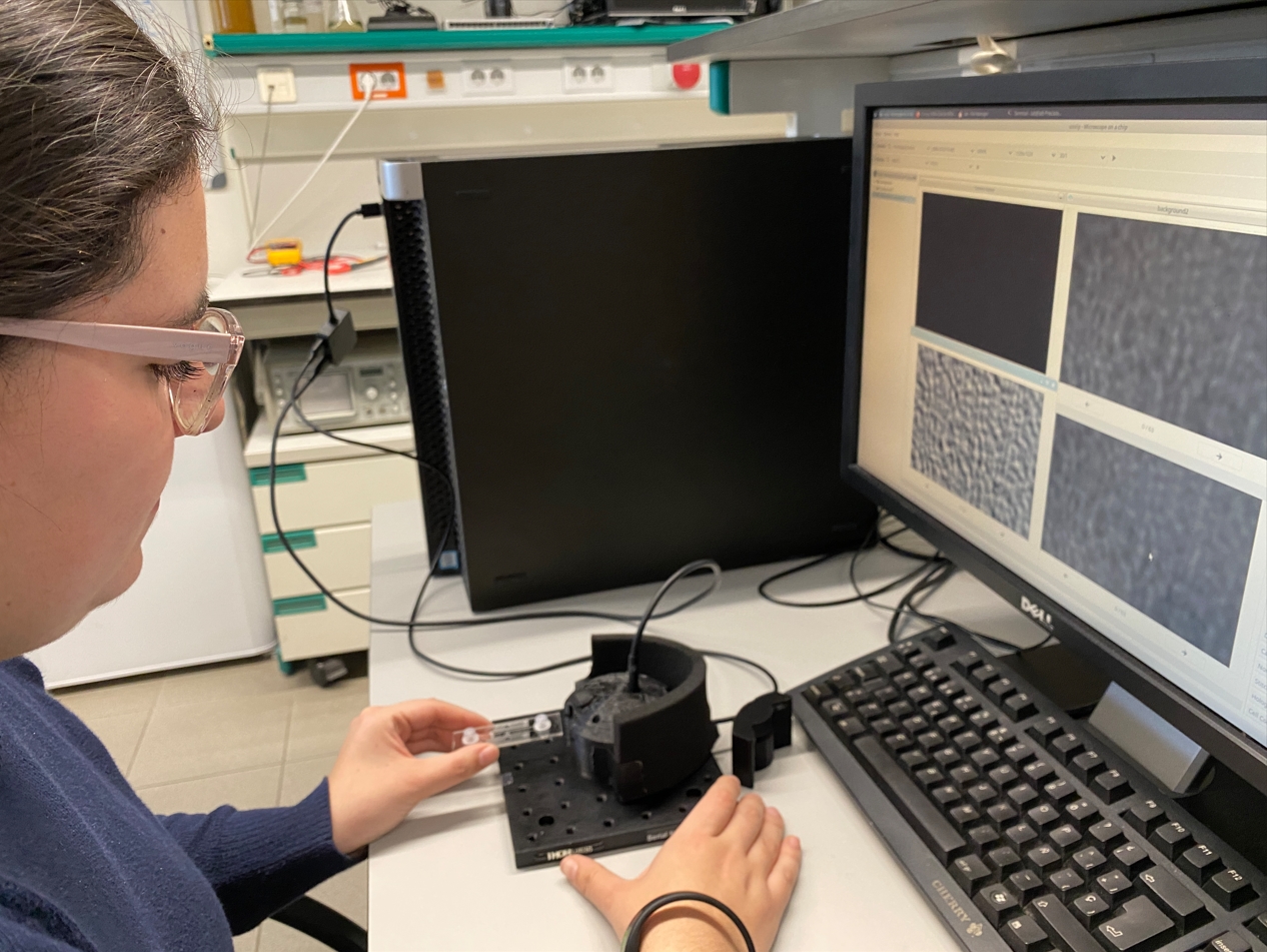
An ultra-compact microscope for monitoring organs on a chip
Microscopic measurements are traditionally performed by placing the sample on the microscope stage. Now, researchers at the University of Barcelona (UB) have developed an ultra-compact device that allows the instrument to be brought to the sample and, therefore, analyzed continuously and in real time.
The project, led by the professor of the Department of Electronic and Biomedical Engineering of the Faculty of Physics and member of the Institute of Nanoscience and Nanotechnology of the UB (IN2UB), Ángel Diéguez, has received a grant Product of the call Industry of Knowledge 2023 of the Generalitat of Catalonia of 150,000 euros to produce a prototype of this innovative technology. It is expected that the microscope will have applications in different areas, from biomedical research or the development of drugs, to the control of water quality or the wine production process.
The microscope is based on a CMOS camera – a technology for the construction of integrated circuits that allows for great miniaturization – and a micro-screen with small LEDs that illuminate the object to create shadow images of the sample. These images are captured with a very sensitive detector and processed with artificial vision algorithms to extract information relevant to the user.
A technology to revolutionize biomedical research
The goal of the researchers in this phase of the technology is to use this microscope to monitor organs on a chip (OOC, acronym for the English word Organ-on-a-Chip), an emerging technology that, in the words of Professor Diéguez, is “revolutionizing the way biomedical research is carried out”, and that this project is being developed by researchers from the same UB department led by Professor Oscar Castaño.
It is a cell culture methodology that, using computer-controlled chips and sensors that measure cell metabolism, simulates the microenvironment and key functional aspects with the aim of mimicking the behavior of a living organ on a microscopic scale. “In this way, an in vitro environment is achieved, which can be easily replicated, drastically speeding up biomedical tests and the discovery of new drugs, saving the use of animals in this type of experiments”, explains the UB researcher.
Simplification and automation of experiments
One of the obstacles to the use of these bodies is their monitoring, especially for conducting mass trials, which need to simultaneously monitor large sets of OOC devices. Despite advances in automated image processing and interpretation algorithms, even today the evolution of tissues can only be monitored manually, by taking them one by one to a conventional microscope for observation.
“Our portable microscope with a size of a few millimeters is a perfect solution to this problem, since a device can be installed in each organ on a chip for continuous and automated monitoring, reducing the effort, materials and costs of the essays”, explains Ángel Diéguez.
According to the researcher, this new microscope would “drastically simplify” the performance of this type of biomedical tests, also improving “the quality of the information collected, thanks to the continuity, completeness and lack of external manipulation of the samples”.
In the new project, docket number 2023 PROD 00008, the researchers will develop a prototype of their miniature microscope and integrate it into an organ-on-a-chip, also incorporating signal processing technology to demonstrate the functionality and utility of the concept They will also work on the creation of a spin-off company to carry out the process of industrialization and commercialization of this unique microscope.
These grants from the Generalitat’s Knowledge Industry program are intended for obtaining prototypes and for the valorization and transfer of research results generated by research teams in Catalonia.

