

Microscopes-On-a-Chip (MOC)
Advantages
- Continuous real-time data analysis for rapid decision making.
- Parallel processing of multiple samples to improve efficiency and resources.
- Autonomous system operation, error reduction through automation.
- Cost-effective prototyping.
- Multiple imaging techniques for application in various fields of life sciences.
Goal
Industrial partners are being sought to commercially exploit the technology.
Patent
International Patent Application
Priority date: January 2021
Currently at national phases in EU and US
Reference
UBTT0401b
Contact
Dr. Sancho Moro
Email: smoro@fbg.ub.edu
Tel: +34 673388625
Microscopes-On-a-Chip (MOC)
Executive summary
A microscope-on-a-chip (MOC) device based on microelectronic technologies has been developed by researchers from the University of Barcelona. The compact microscope is attached directly to the sample, contrary to the conventional microscopes where the sample is brought to it. In addition, our device allows real-time monitoring, simultaneous analysis, and autonomous recognitions methods in the specimen culture conditions.
Introduction
Ultra-compact microscopes can be used in place of conventional microscopes in scenarios that require a smaller instrument size, simultaneous multiple sampling, and cost-effectiveness. In traditional microscopy, the sample must be brought close to the plate, but microscopes of such small size can be placed around the specimen. Real-time monitoring, parallel data acquisition, and minimal direct user intervention are other key attributes of these ultra-compact microscopes. Their direct application in the healthcare sector could be particularly advantageous. For example, in drug development processes, Organ-On-a-Chip (OOC) technology is currently being tested as a substitute for animal testing to speed up the drug discovery process. Our ultra-compact microscopes can be attached to each OOC in the experiment, eliminating the need to transport samples from the incubator to the microscope. This eliminates the need to open the incubator, thus avoiding loss of conditioning, and simultaneously reduces the consumables needed for the procedure.
Description
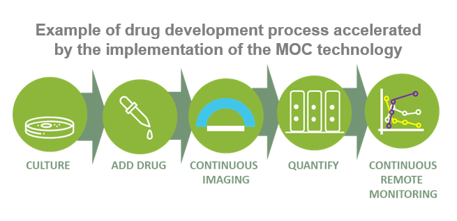
The prototype of the microscope is based on a CMOS camera and a microdisplay with LED technology. The use of various imaging modes, activated by arbitrary LEDs on the microdisplay, allows for versatile and precise observation. In addition, the compact device employs advanced vision algorithms to process the reconstructed images, extracting relevant information for the user. Figure 1 shows an example of the use of MOC in a drug development process. Thanks to the integration of the microscope with the samples, they can be monitored continuously. Therefore, sample analysis is accelerated by parallelizing the process. In addition, the analysis of the sample takes place without the need for user intervention.
Current stage of development
A working prototype at the TRL4-5 has been developed, expecting that to be demanded and adopted by end-users.